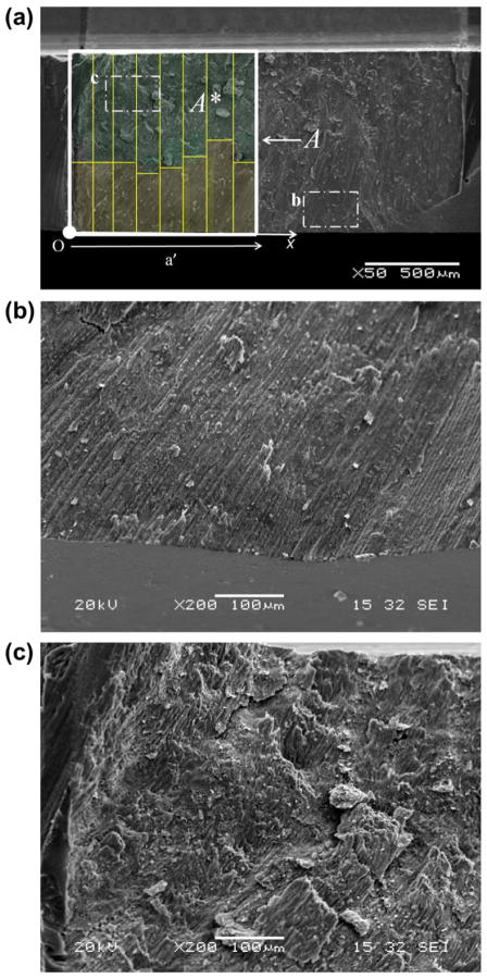
Fig. 2.
Characterizing the degree of decussation from the fracture surfaces. The direction of crack extension is from left to right. (a) SEM micrograph of a fracture surface. The rods uniformly extend from the occlusal surface (bottom) but begin to undergo decussation close to the back channel. The degree of decussation at a specific crack length is defined as the ratio of decussated fracture area A* to total fracture area A. (b) Micrograph of non-decussated outer enamel close to occlusal surface. (c) Micrograph of heavily decussated inner enamel close to back channel.

