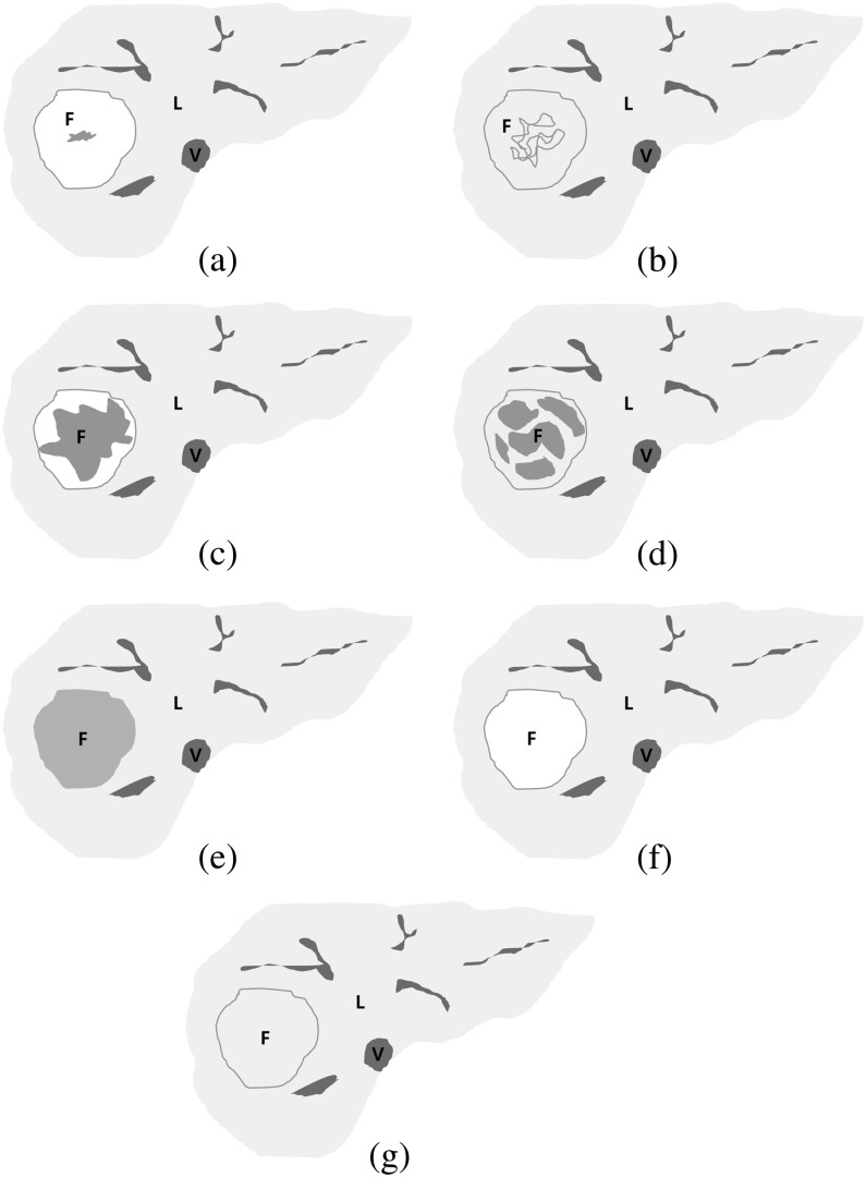
Figure 1.
Schematic diagrams of focal nodular hyperplasia (FNH) on hepatobiliary phase images of gadoxetic acid-enhanced MRI. (a) Heterogeneous high signal intensity (SI), (b) heterogeneous iso SI, (c) heterogeneous low SI, (d) heterogeneous low SI (2), (e) homogeneous low SI, (f) homogeneous high SI, (g) homogeneous iso SI. Heterogeneous high, iso or low signal intensity (SI) lesions and homogeneous low SI lesions were categorised into the fibrosis group (a–e), and homogeneous high or iso SI lesions were categorised into the non-fibrosis group (f, g). F, FNH; L, liver; V, inferior vena cava.

