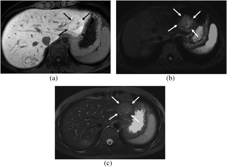
Figure 2.
A 32-year-old female. (a) Hepatobiliary phase image of gadoxetic acid-enhanced MRI shows a focal nodular hyperplasia showing heterogeneous high signal intensity (SI) (arrows), which is the fibrosis group. (b) On diffusion-weighted image (b=400), the lesion shows high SI (arrows) and diffusion restriction. (c) On T2 weighted image with fat suppression, the lesion shows high SI compared with that of the hepatic parenchyma (arrows).

