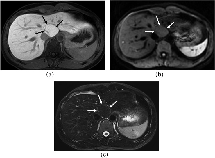
Figure 6.
A 29-year-old female. (a) Hepatobiliary phase image of gadoxetic acid-enhanced MRI shows a focal nodular hyperplasia showing homogeneous iso signal intensity (SI) (arrows), which is the non-fibrosis group. (b) On diffusion-weighted image (b=400), the lesion shows iso SI (arrows) and lacks diffusion restriction. (c) On T2 weighted image with fat suppression, the lesion shows iso SI compared with that of the hepatic parenchyma (arrows).

