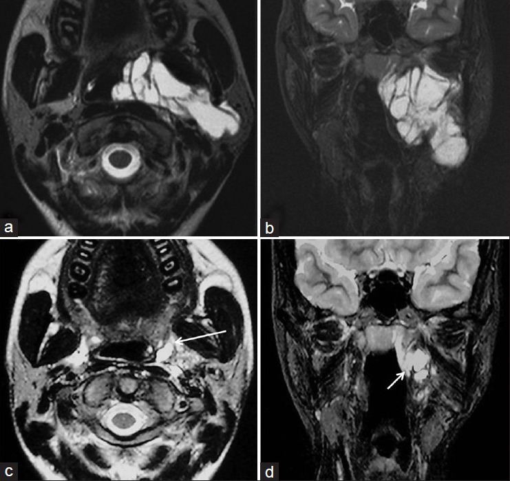
Figure 5.

A 16-year-female with lymphatic malformation in the left parapharyngeal space treated with Bleomycin sclerotherapy; (a-b) Axial and coronal T2 weighted images showing the hyperintense lymphatic malformation; (c-d) Axial and coronal T2 weighted images after one session of percutaneous aspiration and Bleomycin sclerotherapy showing the residual lymphatic malformation (arrows)
