
Figure 4.
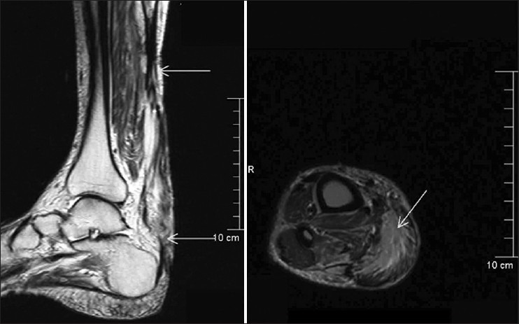
Magnetic resonance imaging T2-image showing the tendon defect, which is filled by the muscle graft and the deeper part of the muscle graft is in continuity with the proximal myotendinous region and distal Achilles tendon – suggestive of complete graft incorporation