
Figure 9.
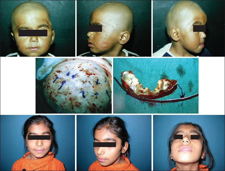
A plagiocephaly patient (upper row) showing clinical features. The middle row shows fused coronal suture on one side. The fronto-orbital segment shows recessed segment on affected side. The post-operative appearance at 6 years later (lower row)