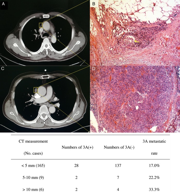
Figure 1:
(A and B) A false-negative case on CT scan. A tiny Station 3A node (A) turned out to be tumour metastasis in the fatty tissue (B). These tumour cells formed loci within a lymph vessel (haematoxylin–eosin, original magnification, ×200, arrow). (C and D) shows a false positive case of CT scan. A distinctively enlarged Station 3A node (C 13 mm at short-axis diameter) was actually lymphoid hyperplasia in histopathological section (D haematoxylin–eosin, original magnification, ×100). Early signs of tumour cell implant into lymph nodes were noticed in 4 cases, manifesting in histology as tumour cells located only within the cortex of lymph node or lymph vessels.

