FIGURE 2.
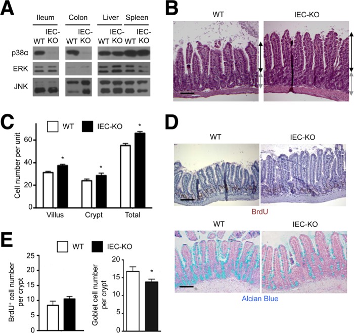
Epithelial-specific p38α ablation leads to a proliferation-differentiation imbalance in the intestinal mucosa. A, whole cell lysates from WT and IEC-KO tissues were analyzed by immunoblotting with antibodies against the proteins indicated on the left. B–E, ileum tissue sections from WT and IEC-KO mice were analyzed by hematoxylin and eosin staining (B), immunostaining with BrdU-specific antibody (D, upper panel), and Alcian Blue staining (D, lower panel). Black and gray lines with double arrows indicate villus and crypt length, respectively (B). Scale bar, 100 μm. The numbers of epithelial cells constituting villus and crypt units (C), and the numbers of BrdU+ cells and Alcian Blue+ goblet cells per crypt (E) were determined. Data represent means ± S.E. *, p < 0.05.

