FIGURE 4.
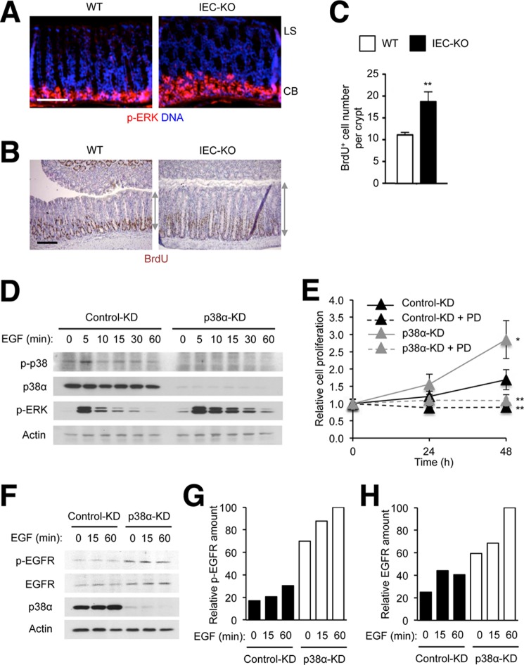
Intestinal epithelial cells lacking p38α exhibit ERK hyperactivation at the crypt base and proliferate at higher rates in response to EGF. A–C, colon tissue sections from WT and IEC-KO mice were analyzed by immunostaining with p-ERK- and BrdU-specific antibodies (A and B, respectively). LS, luminal surface; CB, crypt base. Gray lines with double arrows indicate crypt length (B). Scale bar, 100 μm. The number of BrdU+ cells per crypt was determined (C). Data represent means ± S.E. **, p < 0.01. D–H, MODE-K cells were transfected with siRNA as described in Fig. 1 and treated with EGF (10 ng/ml) and PD98059 (PD; 20 μm) as indicated. After addition to the cells, EGF was present in the culture media throughout the incubation period. Whole cell lysates were prepared after the indicated durations of stimulation and analyzed by immunoblotting with antibodies against the proteins indicated on the left (D and F). Cell proliferation (E) and p-EGFR and EGFR amounts (G and H) were determined at the indicated time points after stimulation. The relative protein amount denotes the percentage of the protein intensity relative to the maximum in each immunoblot. Data represent means ± S.E. **, p < 0.01 (PD98059-treated versus untreated); *, p < 0.05 (p38α-KD versus control KD).

