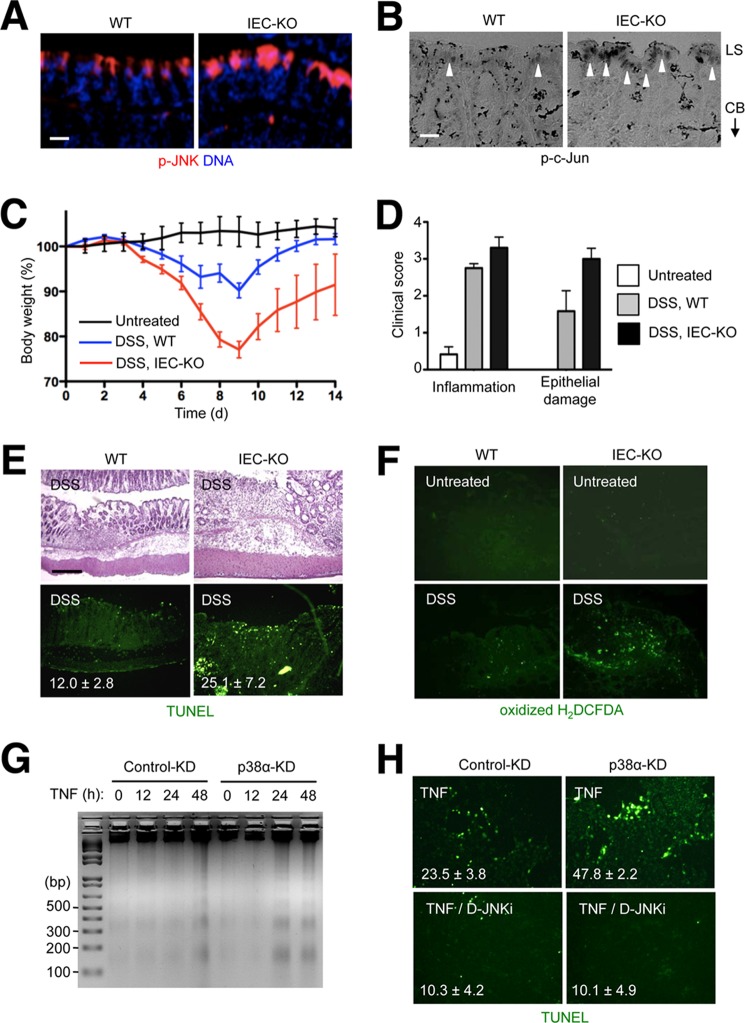
FIGURE 5.
Intestinal epithelial cells lacking p38α exhibit JNK hyperactivation at the luminal surface and predispose to inflammation-induced apoptosis. A and B, colon tissue sections from WT and IEC-KO mice were analyzed by immunostaining with p-JNK- and p-c-Jun-specific antibodies (A and B, respectively). LS, luminal surface; CB, crypt base. Arrowheads indicate areas where specific immunostaining is detected. Scale bar, 10 μm. C–F, WT and IEC-KO mice were orally administered DSS (3.5% in the drinking water) for 7 days. Changes in body weight were monitored daily (C). Colonic tissue sections were prepared on day 7 and analyzed by hematoxylin and eosin staining and TUNEL staining (E, upper and lower panels, respectively). Colonic tissue sections from untreated and DSS-treated mice were incubated with 2′,7′-dichlorodihydrofluorescein diacetate (H2DCFDA) and analyzed by fluorescence microscopy (F). Clinical scores indicate the extent of inflammation (leukocyte infiltration and edema) and epithelial damage (D). Data represent means ± S.E. G and H, MODE-K cells were transfected with siRNA and treated with TNF (10 ng/ml) as described in Fig. 1 in the absence and presence of D-JNKi (1 μm). The rate of apoptosis was analyzed by agarose gel electrophoresis and ethidium bromide staining of genomic DNA obtained from the cells at the indicated time points (G) and TUNEL staining 16 h after TNF treatment (H). The number within each image indicates percentage of TUNEL+ cells and represents mean ± S.D.

