FIGURE 6.
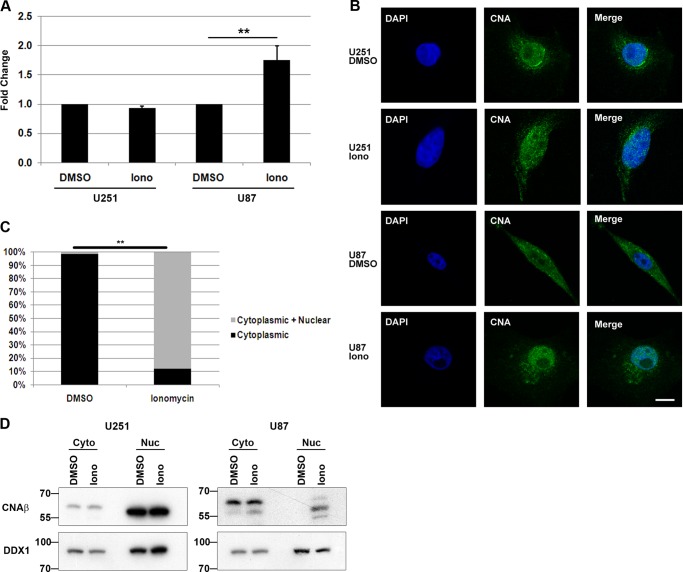
Ionomycin alters NFI-dependent promoter activity and calcineurin localization. A, U251 and U87 cells were transfected with pCAT/GFAP and treated with 1 μm ionomycin (Iono) or DMSO (control) for 24 h. Acetylated [14C]chloramphenicol was measured in counts/min from equal aliquots of cell lysates using a scintillation counter. The fold increases in CAT activity are relative to the DMSO control. The results are an average of four independent experiments with standard deviation indicated by error bars. B, subcellular localization of calcineurin in U251 and U87 cells treated with DMSO (control) or 1 μm ionomycin (Iono) was analyzed by immunofluorescence using anti-CNA primary antibody followed by Alexa 488-conjugated secondary antibody. DNA was stained with 4′6-diamidino-2-phenylindole (DAPI). Bar, 10 μm. C, percentage of cells with predominantly cytoplasmic staining versus cells with nuclear and cytoplasmic staining for CNA in U87 cells treated with DMSO (control) or 1 μm ionomycin for 1 h. This analysis was carried out >100 cells for each parameter. Briefly, 10 separate fields with ∼10–15 cells per field were randomly selected for each parameter. Line scans through the cytoplasm and nucleus of each cell were used to assess relative signal in the nucleus and cytoplasm. Statistical significance was determined using unpaired t test. **, denotes p < 0.001. D, U251 and U87 cells were treated with DMSO (control) or 1 μm ionomycin (Iono) for 1 h and harvested, and cytoplasmic (Cyto) and nuclear (Nuc) extracts were prepared. Extracts were electrophoresed, transferred to PVDF membranes, and immunostained with anti-CNAβ and anti-DDX1 (loading control) antibodies.

