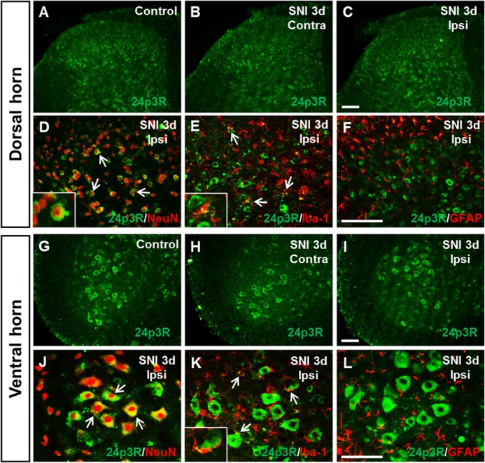
FIGURE 6.

Immunolocalization of 24p3R (the LCN2 receptor) in spinal cord after SNI. A–C and G–I, 24p3R immunoreactivity in naive control animals (A and G), in the contralateral (Contra) dorsal and ventral horn of the spinal cord (B and H) and in the ipsilateral (Ipsi) dorsal and ventral horn (C and I) of the spinal cord at 3 days after SNI. Scale bars, 100 μm. D–F and J–L, double immunostaining showed that 24p3R (green) in the dorsal (D--F) and ventral horn (J–L) of the spinal cord was co-localized with NeuN (red) or Iba-1 (red), but not with GFAP (red). High magnification images (insets in D, E, and K) indicate doubly labeled cells in the ipsilateral dorsal (D and E) or ventral horn (K), respectively. Arrows indicate examples of doubly labeled cells. Scale bars, 100 μm. Results are representative of more than three independent experiments.
