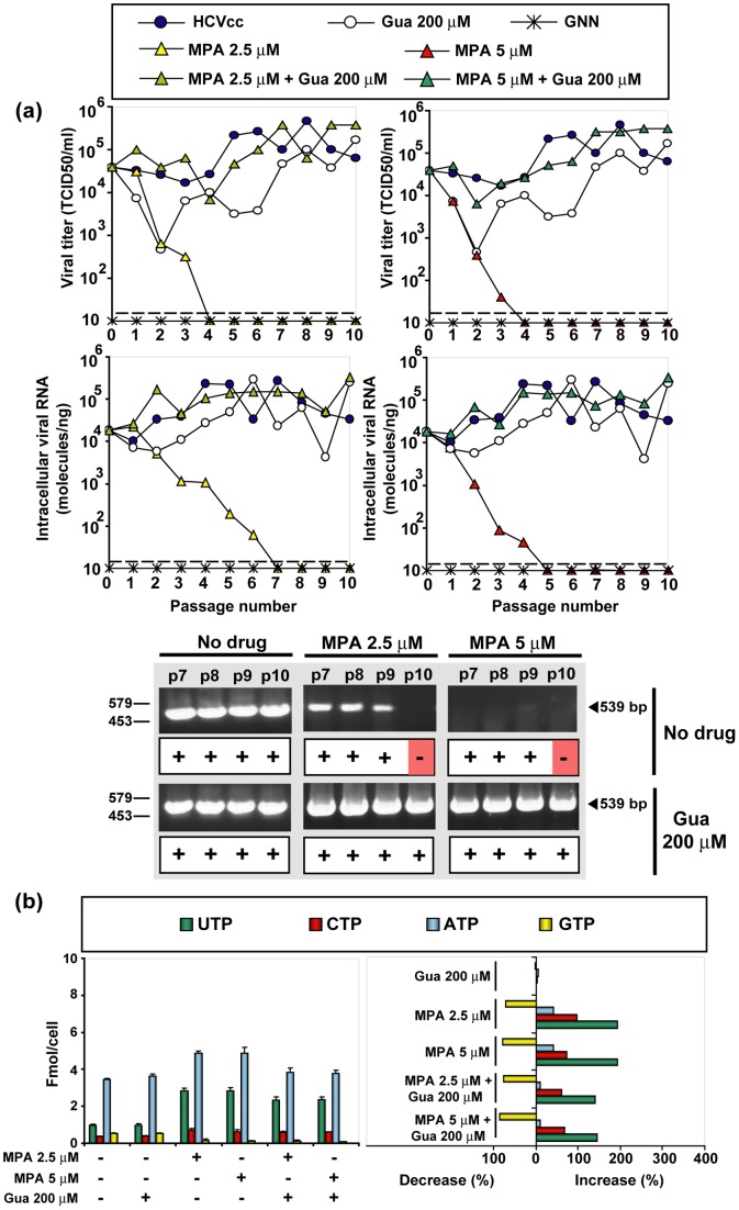
Figure 6. The effect of mycophenolic acid (MPA) and guanosine (Gua) on HCV infectious progeny production and intracellular nucleotide pools.
(a) Huh-7.5 reporter cells were infected with HCVp0 at an initial MOI of 0.1–0.2 TCID50/cell, in the absence or presence of the MPA and Gua concentrations indicated in the upper box. Infections with HCV GNN were carried out in parallel (negative control). The progeny form each infection was used to infect fresh cells, as described in Materials and Methods. Viral infectivity was determined in the cell culture supernatant (upper panels), and viral RNA quantitative RT-PCR in an extract of infected cells (lower panels). The discontinuous lines parallel to the abscissa indicates the limits of detection of infectivity and viral RNA. Below, RT-PCR amplification bands using a highly sensitive HCV-specific amplification protocol that yields a 539 bp fragment, using as template total intracellular RNA from the infection series and passage numbers indicated at the top of the corresponding lanes; +, −: presence or absence of amplification band. (b) Left: Intracellular amount of the four nucleoside-triphosphates, following 72 h of exposure to either MPA or Gua, as indicated at the bottom. Right: decrease or increase of nucleotide concentration as a result of exposure to Gua or MPA; note that the maximum decrease possible is 100%. Procedures are described in Materials and Methods.

