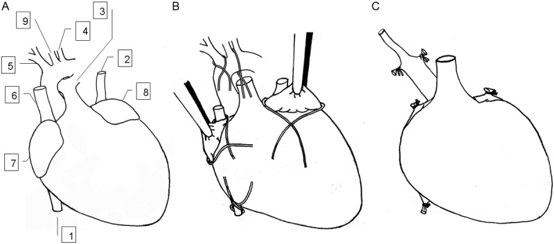
Figure 1.

Paradigm for harvesting cardiac graft. A: In situ location of blood vessels in the donor heart. B: Ligation placed on the donor heart. The inferior vena cava was ligated, the right superior vena cava was ligated with homolateral auricular appendix and dissected distally. The left auricular appendix was ligated and dissected distally. The innominate artery and the aortic arch between the left common carotid artery and left subclavian artery were ligated and divided distally. The left common carotid artery was isolated from surrounding fat tissues and transected as distally as possible. The pulmonary artery was transected at its first bifurcation. C: The prepared cardiac graft ready for implantation. 1, inferior vene cava; 2, left superior vena cava; 3, pulmonary artery; 4, left subclavian artery; 5, innominate artery; 6, right superior vena cava; 7, right auricular appendix; 8, left auricular appendix; and 9, left common carotid artery.
