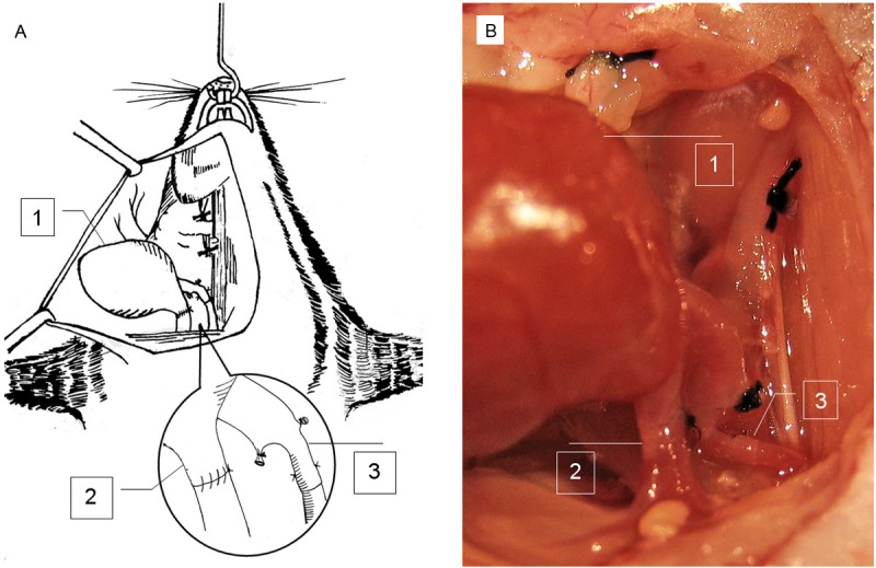
Figure 3.

Schematic view of cervical cardiac implantation. A: A graphic picture for clear illustration of cervical cardiac transplantation. B: An image of transplanted cardiac graft. The submandibular gland is ligated and removed. Venous anastomosis is completed by one-suture end-to-end microsuture technique. Arterial anastomosis is finished by the two-stitch sleeve technique. The inset shows the arterial and venous anastomosis. 1, cardiac graft; 2, the donor pulmonary artery is anastomosed to the recipient right external jugular vein in an end-to-end manner; and 3, the donor right common carotid artery is anastomosed to the recipient right common carotid artery by the end-in-end sleeve technique.
