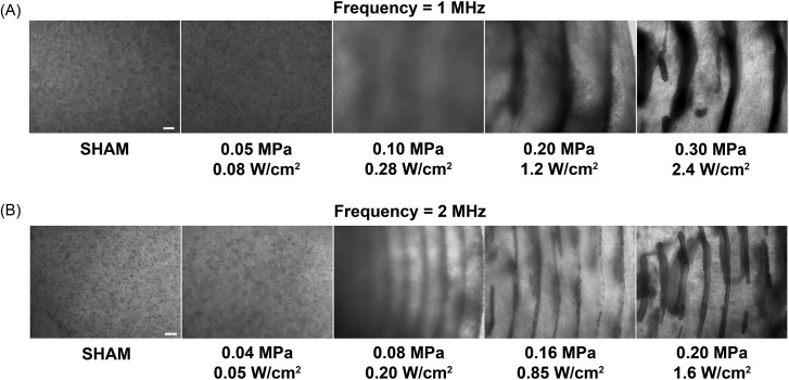
Figure 1.
Spatial patterning of cells within collagen hydrogels using ultrasound standing wave fields. Unpolymerized solutions of type I collagen and endothelial cells (4 × 10⁶ cell/ml) were exposed for 15 min during the polymerization process to either a 1- or 2-MHz CW ultrasound standing wave field. Samples were exposed to various peak positive pressure amplitudes (measured at standing wave field pressure antinodes) and calculated Ispta values. Resultant collagen gels were analyzed for cell location using phase-contrast microscopy. Representative side-view images of cell-embedded collagen gels are shown for 1-MHz (A) and 2-MHz (B) experiments (n = 3). Scale bar, 200 μm.

