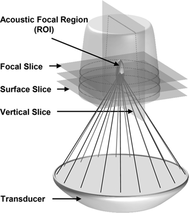
Figure 2.
Schematic representation of the AAP position above the HIFU transducer. The acoustic focus was positioned directly in the center of the phantom. Horizontal MR slices were collected at the focus, at the surface of the phantom, and in between these two slices. During separate scans, vertical slices were collected parallel to the axial length of the acoustic focus.

