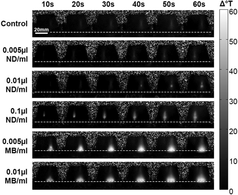
Figure 7.
MR thermometry maps of phantoms containing no agents (top row), ND (second to fourth rows) or MB (fifth and sixth rows). Frames shown were collected during the 60 s of HIFU ablation and indicate the intensity and location of the temperature change. The dotted line across each series of frames delineates the surface of the phantoms.

