Figure 2.
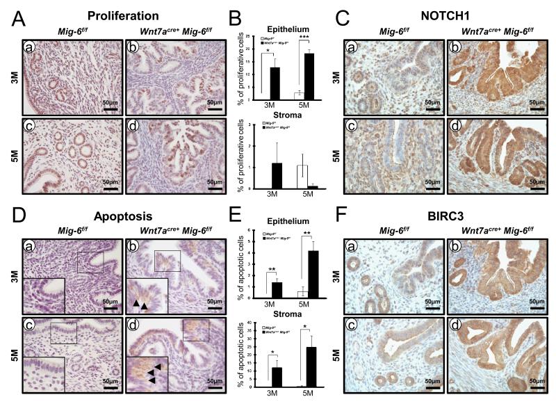
Regulation of proliferation and apoptosis by epithelial Mig-6 (A) Immunostaining of phospho-histone H3 was significantly increased in the endometrial epithelial cells of Wnt7acre+ Mig-6f/f mice (b and d) compared to control mice (a and c) at three months of age (a and b) and five months of age (c and d). (B) Quantification of phospho-histone H3-positive in endometrial stroma and epithelial cells. (C) Immunohistochemical analysis of NOTCH1 in the uteri of control mice (a and c) and Wnt7acre+ Mig-6f/f mice (b and d) at three months of age (a and b) and five months of age (c and d). (D) Immunohistochemistry of cleaved caspase-3 was increased in epithelial cells and sub-epithelial storma cells of Wnt7acre+ Mig-6f/f mice (b and d) compared to control mice (a and c) at three months of age (a and b) and five months of age (c and d). (E) Quantification of cleaved caspase-3-positive in endometrial stroma and epithelial cells. (F) Immunohistochemical analysis of BIRC3 in the uteri of control mice (a and c) and Wnt7acre+ Mig-6f/f mice (b and d) at three months of age (a and b) and five months of age (c and d). Arrowheads indicate positive-cleaved caspase-3 cells. The results represent the mean ± SE. *, p < 0.05; **, p < 0.01; ***, p < 0.001.

