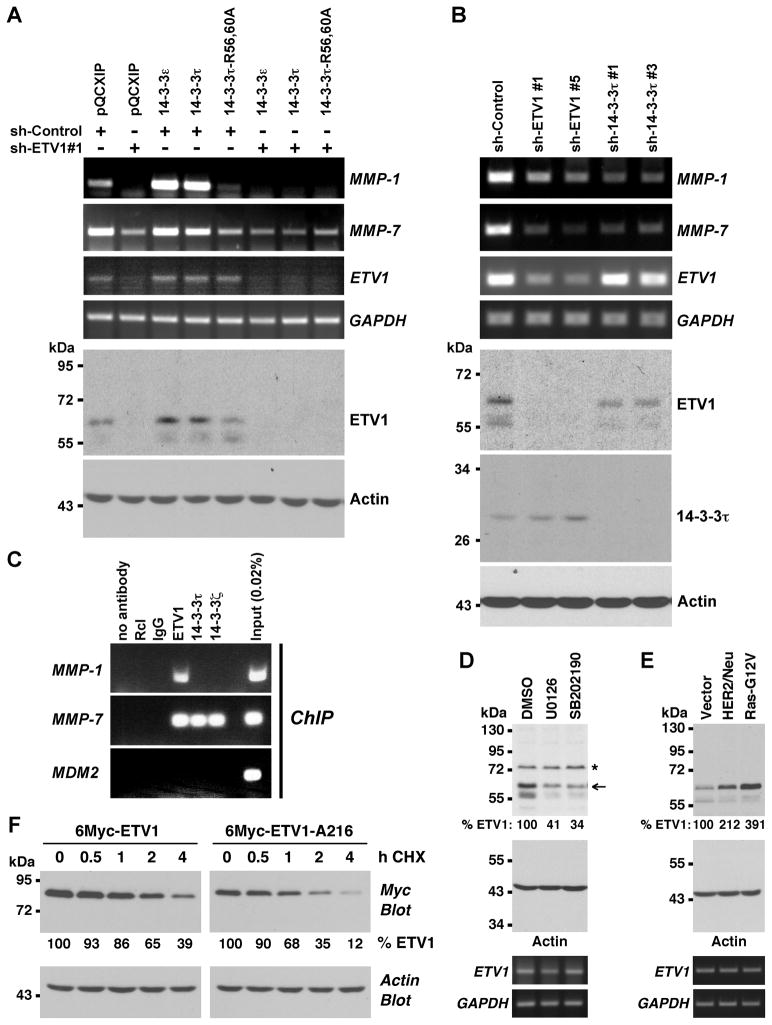
Figure 3.
14-3-3 proteins affect ETV1 activity and stability in prostate cancer cells. (A) LNCaP cells were infected with retrovirus expressing control shRNA or ETV1 shRNA. In addition, cells were infected with retrovirus expressing indicated 14-3-3 proteins or empty vector pQCXIP. MMP-1, MMP-7, ETV1 and GAPDH mRNA levels were assessed by RT-PCR. The bottom two panels show Western blots of ETV1 or actin. (B) Similar analysis of LNCaP cells infected with retrovirus encoding ETV1 or 14-3-3τ shRNAs. (C) ChIP assays in LNCaP cells utilizing indicated antibodies for precipitation. MMP-1 and MMP-7 promoter regions encompassing ETV1 binding sites were amplified, as was the control MDM2 promoter. Rcl antibodies, IgG and no antibody served as negative controls. (D) LNCaP cells were treated with 10 μM U0126 or 5 μM SB202190 24 h and 12 h before lysis and ETV1 levels (normalized to actin) determined by Western blotting. Asterisk marks an unspecific band and arrow points at ETV1. Bottom two panels show corresponding RT-PCR measurements of ETV1 and GAPDH mRNA. (E) Retrovirus encoding HER2/Neu or Ras-G12V was employed to infect LNCaP cells and ETV1 protein and mRNA levels were determined. (F) 6Myc-tagged ETV1 or its A216 mutant was expressed in LNCaP cells. Protein synthesis was then inhibited with 50 μg/ml cycloheximide (CHX) and ETV1 protein levels measured for up to 4 h after the addition of CHX by anti-Myc Western blotting. Relative ETV1 protein amount normalized to actin is indicated.

