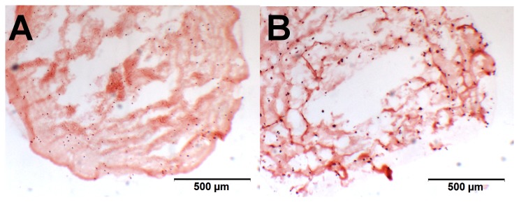
Figure 10. Alizarin red background stain in alginate beads.

Freshly prepared beads, stained with AR with (A) representing a bead seeded with chondrocytes and (B) a bead with MSCs. Both show considerable high but very similar background staining, due to the use of CaCl2 for crosslinking. The calcium ions remain crosslinked in the alginate throughout the culture period.
