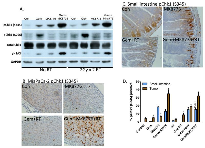
Figure 5. Phosphorylated Chk1 (S345) is a biomarker of MK8776 activity.
A-D, Mice bearing MiaPaCa-2 xenografts (approximately 250mm3) were treated with Gem (120mg/kg, t=0h), MK8776 (50mg/kg, t=2 and 23h), and radiation (2Gy/fraction, t=3 and 24h). One hour post-radiation (t=25h), tumors were lysed for immunoblotting (A) or fixed for immunohistochemistry (B). Small intestines from the same animals were harvested in parallel and prepared for immunohistochemistry (C). D, Quantitation of pChk1 (S345) immunostaining was conducted by scoring the percentage of tumor cells or intestinal crypt cells with nuclear pChk1 (S345) staining. Data are the mean ± standard error of 4 tumors per condition or 2 segments of small intestine from 2 independent animals. Bars represent between 1600 and 3000 scored cells. Statistically significant differences between small intestines and tumor are indicated (*P<0.05). Images/blots are representative of 4 tumors (A, B) or 2 small intestines (C). Complete image sets are included in Suppl. Fig. 5.

