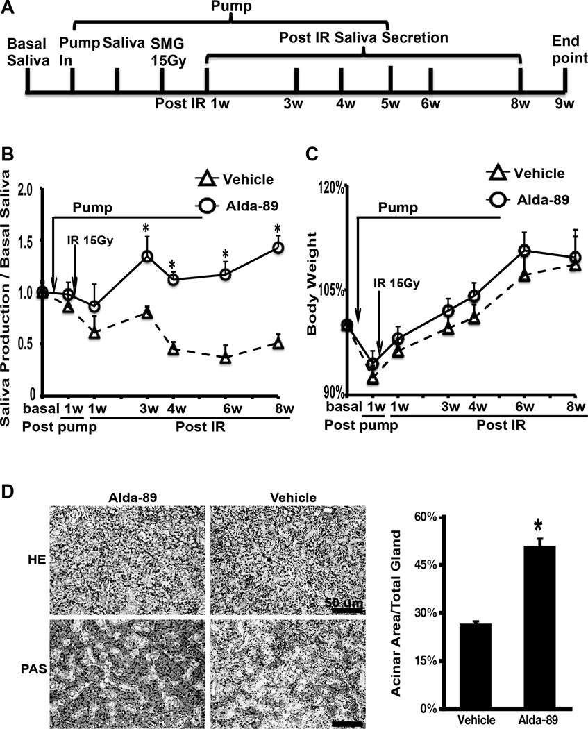
Figure 1. Alda-89 preserves submandibular gland function post radiation.
Panel A:Schematic representation of the experimental procedure. Saliva collection was performed at basal level (before pump placement), 1 week post pump placement, and 1, 3, 4, 6 and 8 weeks post-RT. Mice were euthanized at 9 weeks after RT. Panel B: Whole saliva measurement at different time points by treatment group normalized to the body weight and basal saliva level. Note that the curves diverge around week 3 after RT and the difference was statistitcally significant (* p < 0.05). Panel C: Mean body weight at different time points by treatment group. No significant difference was observed. Panel D: Representive HE staining (top panels, scale bar =100 um) and PAS staining (bottom panels, scale bar = 100um) of SMG tissue showing more intact acinars in the Alda-89 treated glands. Panel E: Quantification of the percent acinar area to total gland area in 10 randomly selected PAS stained images at 200x magnification. There was significantly more intact acini in the Alda-89 treated glands (*p<0.05).

