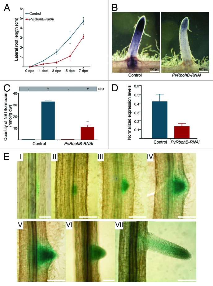
Figure 1. The involvement of PvRbohB in LR growth and development. (A) LR length in control (n = 13, blue line) and PvRbohB-RNAi (n = 15, red line) transgenic roots harvested at 1, 3, 5 and 7 dpe. (B) Detection of superoxide accumulation by NBT (0.1%) staining in control and PvRbohB-RNAi LRs. (C) Quantification of NBT/formazan precipitates per dry weight of LRs stained with NBT (as described by Ramel et al. 200915) as shown in (B). (D) PvRbohB transcript abundance in the apical region (~3 mm from the tip) of control and PvRbohB-RNAi LRs. Expression values were normalized to those of EF1α. Bars indicate means ± SEM from two independent biological replicates (n > 15 independent transgenic roots per biological replicate) with three technical repeats. (E) Promoter activity of PvRbohB, as assessed by bright field microscopy of transgenic roots expressing 1.8 kb of the PvRbohB promoter region fused to GUS and GFP (pPvRbohB:GUS-GFP). Promoter activity was detected by GUS staining during different stages of LR development in P. vulgaris roots (I–VII; n = 18). Scale bar, 100 µm.

An official website of the United States government
Here's how you know
Official websites use .gov
A
.gov website belongs to an official
government organization in the United States.
Secure .gov websites use HTTPS
A lock (
) or https:// means you've safely
connected to the .gov website. Share sensitive
information only on official, secure websites.
