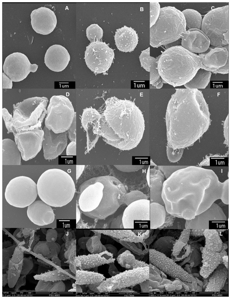
Figure 2. Scanning electron micrographs of test microorganisms with strongly active crude extracts.
Cryptococcus neoformans ATCC 90112 (A–E), Cryptococcus neoformans ATCC 90113 (F), Candida albicans NCPF 3153 (G–I) and a clinical isolate of Microsporum gypseum (K–L) after incubation with 10% DMSO (A, G and J), amphotericin B (B and H), miconazole (K), hexane extract from the mycelia of Penicillium sp. PSU-ES43 (C), hexane extract from the mycelia of PSU-ES190 (D), ethyl acetate extract from the mycelia of Fusarium sp. PSU-ES73 (E and F), ethyl acetate extract from the mycelia of Trichoderma sp. PSU-ES38 (I), and hexane extract from the mycelia of Hypocreales sp. PSU-ES26 (L) for 16 h at 4 times their MIC values.

