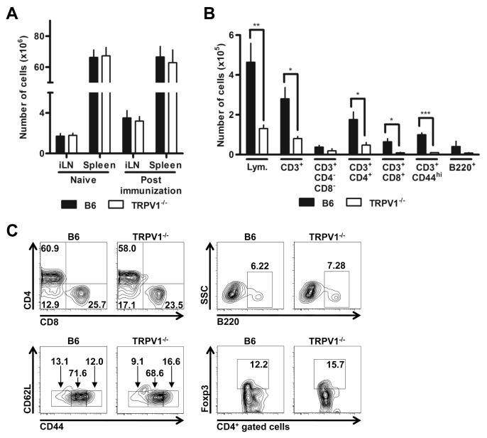
Figure 3.
TRPV1−/− mice show reduced CNS infiltration. (A) Cell counts of inguinal lymph node and spleen prior to and following MOG35–55 peptide immunization of B6 and TRPV1−/− mice (n = 9 per group). (B) Quantification of lymphocytes in the CNS by flow cytometry (n ≥ 3 per group, *p < 0.05, **p < 0.01, ***p < 0.001). (C) Representative FACS plot showing distribution of cells within the CNS; CD4+ and CD8+ cells previously gated on CD3+ cells (upper left), CD62L+ and CD44+ cells previously gated on CD3+ cells (lower left), B220+ cells (upper right), FoxP3+ cells previously gated on CD4+ cells (lower right).

