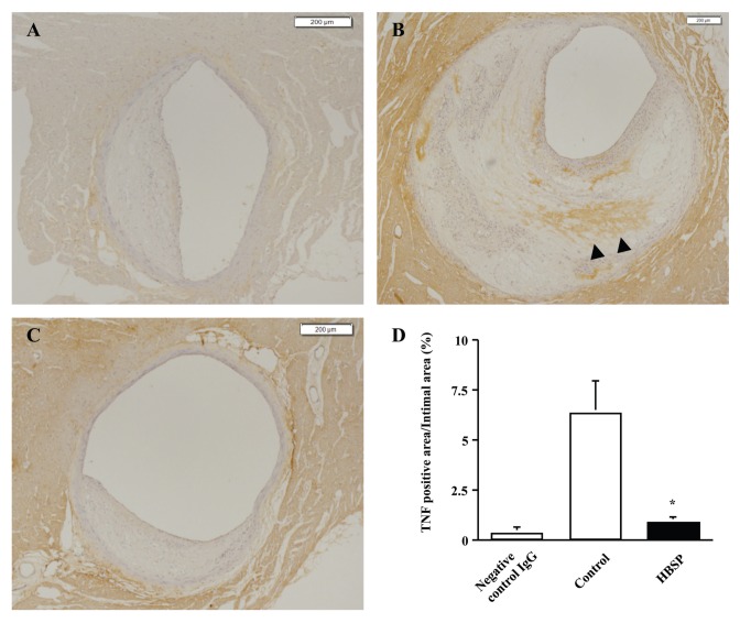
Figure 5.
HBSP reduces TNF-α expression in coronary atherosclerotic lesions of WHHLMI rabbits. (A) Section from control animal immunostained with negative control IgG. (B) Section from control animal immunostained with anti-TNF-α antibody. (C) Section from HBSP-treated animal immunostained with anti-TNF-α antibody. TNF-α–positive areas are visualized in brown (arrowheads). (D) Ratios of TNF-α–positive area/intimal area. Mean ± SEM; negative control IgG, n = 4; control, n = 7; HBSP, n = 8; *P < 0.01. Scale bar, 200 μm.

