Fig. 2.
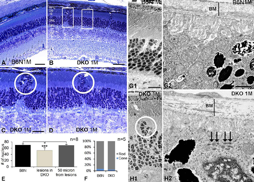
Early focal degeneration of photoreceptors and RPE in DKO mice. A–D: Histological sections in B6N (A) and DKO (B–D) retinas illustrate outward migrating photoreceptors in low (B) and high magnifications (circles in C, D), which was absent in B6N mice (A). E: Histogram shows the number of photoreceptor nuclei in a fixed area (rectangles in B) on B6N and DKO (lesion and 50 µm lateral to the lesion) mice (mean ± SD, **P < 0.001). F: The percentages of cone and rod nuclei in the ONL in B6N and DKO mice, respectively. G, H: EM images show outward migration of photoreceptor nuclei (H1), thickening of Bruch’s membrane (H2, a vertical line) and lipofuscin accumulation in the RPE (H2, arrows) in DKO mice, which are not detected in B6N mice (G1–2). Scale bar: 50 µm for A and B, 20 µm for C and D, 500 nm for G1 and H1, 100 nm for G2 and H2.

