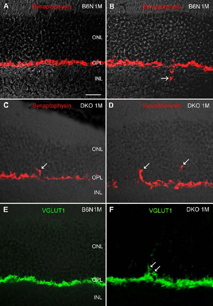
Fig. 4.
Confocal images showing synaptophysin and VGLUT1 IR immunoreactivity on the vertical sections in the B6N and the DKO mice. A, B: Intense synaptophysin IR is always evident in the OPL, but occasionally detectable in the INL (B, arrow) in the B6N mice, indicating inward protrusion of photoreceptor terminals. C, D: In DKO mice, synaptophysin labeled punctas are found in the ONL (arrows). E, F: Similarly, VGLUT1 labeling is found in the OPL in the B6N mice, whereas in the DKO retina VGLUT1 puncta is readily visible in the ONL (F, arrow). Scale bar: 20 µm for A–F.

