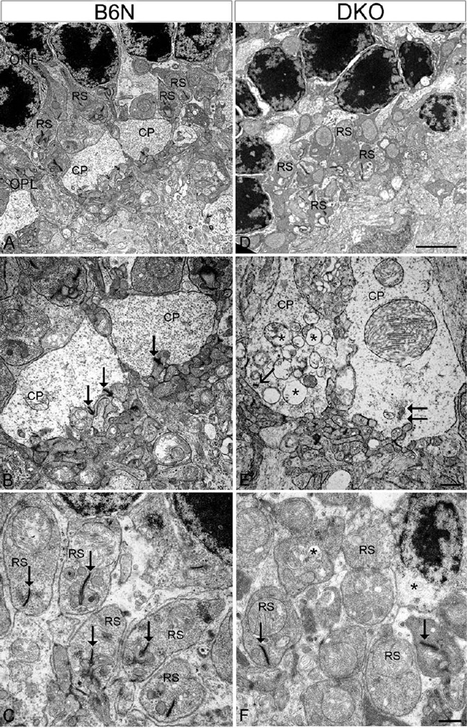
Fig. 6.
EM micrographs showing the degeneration of photoreceptor terminals in DKO mice compared with B6N mice. A–C: In the B6N mice, the cone pedicles (CP) and rod spherules (RS) are organized regularly in the OPL (A), in which CP contains various synaptic ribbons (B, arrows) and RS has only one ribbon (C, arrows). D–F: The DKO mice show highly disorganized photoreceptor terminals in the OPL (D). At high power, DKO retinas show marked reduction of synaptic ribbons and shallower or flattened invaginations in both CP (E) and RS (F). Some CP contain short and “floating” ribbons (E, arrows), but numerous vacuoles (E, asterisks). Synaptic vesicles appear loosely packed in these two degenerate CP (E). However, RS had less vacuole (F) compared with CP. Scale bar: 2 µm for A and D, 500 nm for B, C and E, F.

