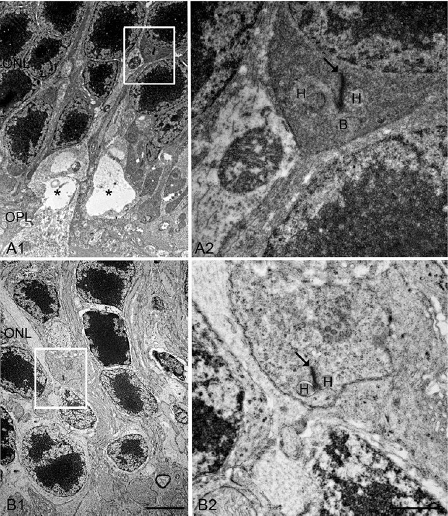
Fig. 7.
EM micrographs showing ectopic rod synapses in the ONL in the DKO mice. A: A rod spherule retracts into the ONL (A1, square), where a new ectopic rod synapse is illustrated at high power image (A2). A similar example is shown respectively on B1 and B2, however, only two HCs are observed in this case. Arrows, ribbons; asterisks, vacuoles; H, HC; B, RBC. Scale bar: 2 µm for A1 and B1, 500 nm for A2 and B2.

