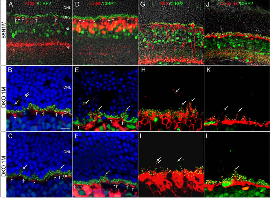
Fig. 8.
Confocal images showing postsynaptic neuron on the vertical sections in the B6N and the DKO mice. A–C: HCN4 IR shows that OFF-CBC dendrites are exclusively in the OPL (vertical arrows) in B6N (A) and DKO mice (B, C), though CtBP2 punctas are visible in the ONL in DKO mice (arrows in B, C). D–F: CaB5 labeled dendrites are exclusively in the OPL of B6N mice (vertical arrows in D) whereas a few dendrites are evident in the ONL in DKO mice (arrows in E, F). G–L: PKC-labeled RBC dendrites (G) and Calbindin-labeled HC processes (J) are restricted in the OPL of B6N retinas. In DKO mice, however, fine PKC-labeled RBC dendrites and Calbindin-labeled HC processes are extended into the ONL, which demonstrate a close association with CtBP2-labeled synaptic ribbons (arrows in H, I, K, and L). Scale bar: 20 µm for A, D, G and J; 10 µm for B and others.

