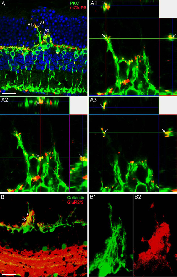
Fig. 9.
Confocal images showing glutamate receptor labeling in the DKO mice. A: mGluR6 puncta are ectopically expressed in the ONL and colocalized with the sprouting of RBC dendrites labeled by PKC (arrows). A1-A3 illustrate three points shown in A through xy, xz, and yz planes (arrows), confirming a colocalization of two proteins. B: Similarly, GluR2/3 puncta are ectopically expressed in the ONL and colocalized with the sprouting of HC processes labeled by Calbindin (arrows). High magnified images are illustrated in Bl and B2. Scale bar: 20 µm.

