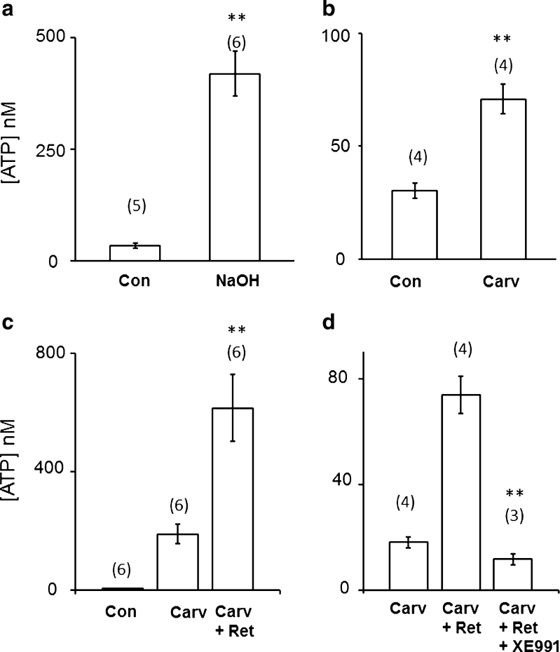
Fig. 7.
Stimulated ATP release from cultured keratinocytes (a, b) and effects of retigabine thereon (c, d). Data in a–d are from four separate keratinocyte cultures. Ordinates show concentrations of ATP ([ATP], in nanomolars) in 50 μl aliquots of Krebs’ solution overlying the keratinocyte cultures (total bath volume 500 μl) determined using the luciferase assay (see “Methods”). Aliquots were taken 10 min after adding 50 μl Krebs’ solution to the keratinocyte chambers (controls, Con) or 10 min after adding: a NaOH (final bath concentration 4 %), b carvacrol (Carv, final bath concentration 1 mM), c 1 mM carvacrol alone or 1 mM carvacrol with 10 μM added retigabine (Ret) and d 1 mM carvacrol, 1 mM carvacrol with 10 μM retigabine and 1 mM carvacrol + 10 μM retigabine 2 min after pre-addition of 10 μM XE991. Numbers in brackets are numbers of culture dishes sampled from each culture; **P < 0.01 (difference from controls or, in d, between carvacrol + retigabine with and without added XE991)

