
figure 1.
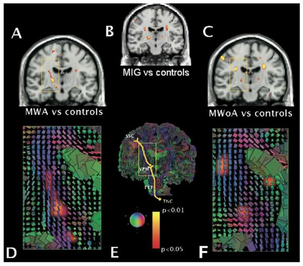
Panels A, B and C show FA average significance maps superimposed on a T1 coronal section comparing migraine with aura (MWA, panel A), all migraine patients (MIG, panel B) and migraine without aura (MWoA, panel C) vs. healthy controls. Panels D, E and F show cuboid maps depicting the diffusion tensor within each voxel. The axes of each cube are placed regarding the main fibres orientation and are coloured accordingly: green indicates antero-posterior fibres; blue indicates supero-inferior fibres; and red indicates medio-lateral fibres. In panels D and F, cuboid maps are superimposed to the FA maps shown in panels A and C, respectively. The trigeminal somatosensory pathway scheme is represented in panel E. The rectangular areas in all panels focus the same anatomical structures. Significant lower FA values were noticed at the thalamocortical tract (3rd order neurons) in both migraine subtypes. In addition, the trigeminothalamic tract (TTT) showed also significantly lower FA values in migraineurs with aura (panel A, lowest cluster inside the yellow square). In both migraine forms, FA clusters were located at the venteroposterior medial thalamic nucleus (VPM), extending upwards along the thalamocortical sensory pathway. The yellow-red shading code represents p values for lower FA changes. n=12 for MWA, MWoA and HC groups. SSC = somatosensory cortex. TNC = trigeminal nucleus caudalis.