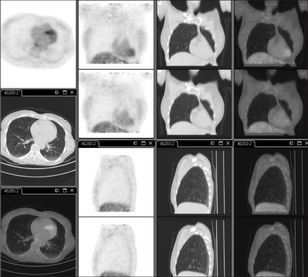
Figure 2.

A control F-18 FDG PET/CT scan performed after 4 days of the first PET/CT. Transaxial-, coronal-, and sagittal-section images of PET alone, CT alone and PET/CT fused images of the lungs revealed complete resolution of the previously noted FDG avid focus. No other abnormalities were noted
