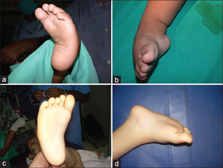
Figure 4.

Clinical photographs showing (a) Plantar and (b) lateral view of the foot showing complete relapse with heel varus, equinus, adduction and deformity. (c) Plantar view of the foot showing adduction deformity of the foot in a 5 year old child. (d) Prone view showing equinus deformity in the same child. These children had Grade III relapse
