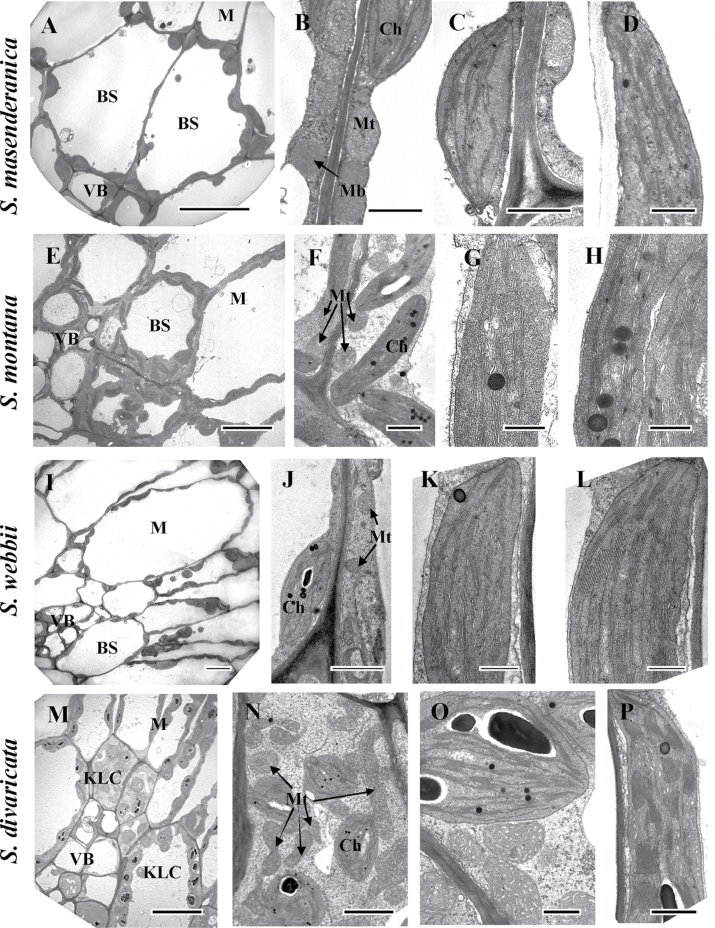
Fig. 4.
Electron microscopy of mesophyll (M) versus bundle sheath (BS) and Kranz-like cells (KLCs) in leaves of four Salsoleae species of formerly Salsola section Coccosalsola: S. masenderanica (A–D), S. montana (E–H), S. webbii (I–L), and S. divaricata (M–P). (A, E, I, M) Micrographs show M and BS/KLCs around vascular bundles. (B, F, J, N, O) Organelles in BS and KLCs at a higher magnification. Note the difference in abundance of organelles in BS and KLCs between species, and the numerous mitochondria in KLCs of S. divaricata (N, O). (C, G, K, O) Chloroplast structure in BS and KCLs of the four species. (D, H, L, P) Structure of M chloroplasts in the four species. Ch, chloroplast; Mb, microbody; Mt, mitochondria; VB, vascular bundle. Scale bars=10 μm for A, E, I, M; 1 μm for B, C, F, J; 0.5 μm for D, G, H, K, L, O, P; and 2 μm for N.

