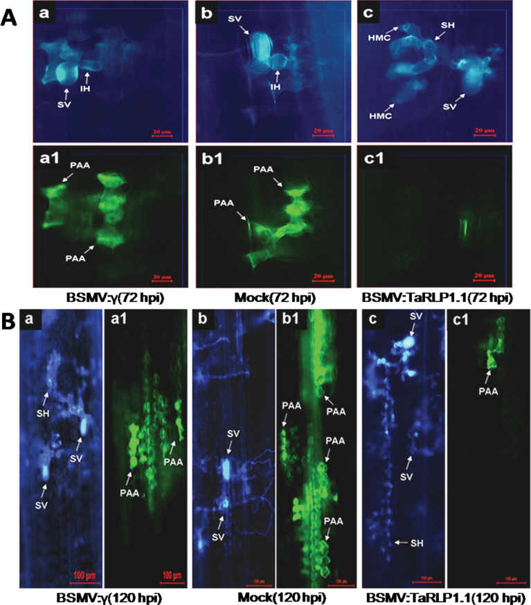
Fig. 4.
Histological observation in wheat cv. 92R137 at 72 hpi (A) and 120 hpi (B) after Pst CYR32 inoculation in the BSMV:γ- (a, a1), mock- (b, b1), and BSMV:TaRLP1.1as- (c, c1) pre-infected leaves. The same infection site was observed by white fluorescence microscopy for fungal growth (a, b, c) and epifluorescence microscopy for hypersensitive cell death (a1, b1, c1). (A) The spreading of secondary hyphae was suppressed (a, b), and mesophyll cells at the infection sites showed strong phenolic autofluorogen accumulation (a1, b1). However, secondary hyphae were formed (c) and autofluorescence was not obvious (c1) in the BSMV:TaRLP1.1as-infected leaves. Bars=20 μm. (B) Secondary hyphae were longer in the BSMV:TaRLP1.1as-infected leaves (c) than in the BSMV:γ- (a) and mock- (b) infected leaves. However, the area of autofluorescence was smaller in the BSMV:TaRLP1.1as-infected leaves (c1) than in the BSMV:γ- (a1) and mock- (b1) infected leaves. Bars=100 μm. SV, substomatal vesicle; IH, infection hypha; HMC, haustorial mother cell; SH, secondary hyphae; PAA, phenolic autofluorogen accumulation.

