Fig. 2.
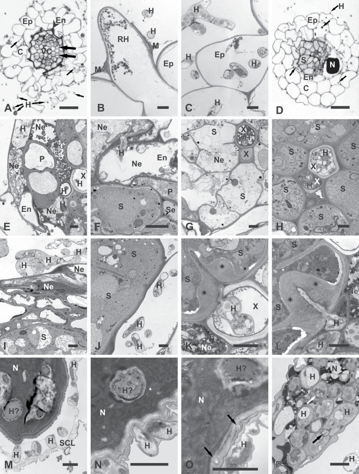
Light (A and D) and transmission electron microscopy (B, C, E–P) micrographs of Arabidopsis roots infected with P. indica only (A–C) or with P. indica and H. schachtii (D–P). (A–C) Cross-sections of roots infected with P. indica (17 dpi; equivalent to 10 days after nematode inoculation): (A) early stage of secondary thickening when cambial stripes had developed (broad arrows) by divisions of procambial cells and endodermis collapsed (arrowhead); fungal hyphae (small arrows) present outside and inside the root; (B) root-hair and epidermal cells with necrotized protoplasts and extracellular hyphae; (C) epidermal cells with necrotized protoplasts and extra- and intracellular hyphae. (D–L) Cross-sections through nematode-induced syncytia. (D–G) At 4 dpi in –7 treatment roots: (D) nematode and syncytium developed inside root infected with hyphae (arrows); (E) hyphae inside necrotized cells of endodermis and the vascular cylinder; some hyphae have entered the lumen of xylem vessels; (F) hyphae inside necrotized endodermal cells next to alive and non-infected syncytium; (G) necrotized syncytium next to vessel invaded with fungal hyphae. (H and I) At 10 dpi in –3 treatment roots: (H) syncytium with regular cytoplasm next to vessel invaded with fungal hyphae; (I) Well-developed network of hyphae separated from syncytium by a layer of cells. (J–L) At 10 dpi in –7 treatment roots: (J) hyphae next to syncytial elements containing regular cytoplasm. (K) hypha growing inside vessel lumen had attempted to penetrate syncytial cell wall that responded with formation of pronounced cell-wall thickening (asterisks); (L) hypha penetrating syncytial cell wall induced formation of pronounced cell-wall thickening (asterisks). (M–P) Nematode bodies and fungal hyphae. (M, O, and P) At 10 dpi in –3 treatment roots: (M) hyphae breaking a subcrystalline layer; (O) possible infection hyphae (arrows) penetrating the cuticle; (P) hyphae inside deteriorated nematode corpse. (N) At 10 dpi in –7 treatment roots: hyphae in direct contact with the cuticle of a juvenile. Arrows indicate septae typical for Sebacinaceae. C, Cortical parenchyma; En, endodermis; Ep, epidermis; H, hypha; H?, hypha-like structure; M, mucilage; N, nematode; Ne, necroses; P, pericycle; RH, root hair; SCL, subcrystalline layer; Se, sieve tube; S, syncytium; X, xylem vessel. Bars: 20 μm (A and D) and 2 μm (B, C, and E–P).

