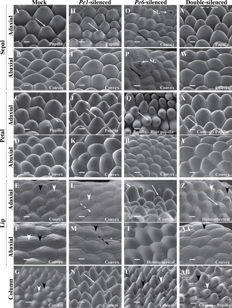
Fig. 6.
Cyro-SEM micrograph of side views of epidermal cells of floral organs of the 7th blooming flowers of mock-treated (A–G), PeMADS1-silenced (H–N), PeMADS6-silenced (O–U), and double-silenced floral organs (V–AB). (A, H, O, V) Sepal adaxial epidermal cells had papilla, papilla, convex, and papilla shapes. (B, I, P, W) Sepal abaxial epidermal cells all had convex shape; St., stomata. (C, J, Q, X) Petal adaxial epidermal cells had papilla, papilla, papilla/hair papilla, and conical/papilla shapes. (D, K, R, Y) Petal abaxial epidermal cells all had convex shapes. (E, L, S, Z) Lip adaxial epidermal cells had convex, convex, conical, and hemispherical shapes. (F, M, T, AA) Lip abaxial epidermal cells had convex, convex, hemispherical, and convex shapes. (G, N, U, AB) Column epidermal cells had conical, conical, conical, and different shapes (including convex, hemispherical cupola, conical, and papilla). Solid white arrows indicate a protuberance on the top of the cell; solid black arrow indicates epicuticular wax crystals; white arrowheads indicate irregular cuticular folds in the central field; black arrowheads indicate parallel cuticular folds in the central field; dashed white arrows indicate irregular cuticular folds in the anticline field; dashed black arrows indicate parallel cuticular folds in the central field. Bars, 25 μm. Materials were Dtps. OX Red Shoe ‘OX1407’.

