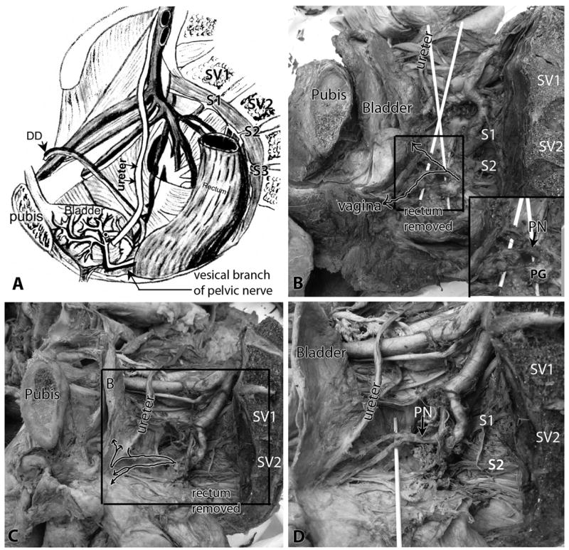
Figure 1.

Identification of the pelvic nerve and its vesical branch to the bladder within the pelvic cavity. Lateral view, with pubis (pubic bone) and sacral vertebrae (SV) indicated as landmarks. A) Diagram of male pelvis showing the relationship of the vesical branch of the pelvic nerve to the bladder, ureter and ductus deferens (DD). B) Pelvic cavity of a female cadaver showing similar relationships. Arrows in the box indicate the path of the pelvic nerve (PN), shown passing over wooden sticks. One branch passes to the bladder (the vesical branch of the pelvic nerve) and a second passes to the vaginal area in this cadaver. The rectum is removed. Inset in B shows enlargement of boxed area, and shows the relationship of the pelvic nerve (PN) with the pelvic ganglia (PG). C) The pelvic cavity of a second cadaver showing similar relationships. Arrows in box indicate the path of the pelvic nerve, and shows one branch passing to the bladder (the vesical branch of the pelvic nerve) that then splits into a branch to the ureter and two branches to the detrusor wall. A second branch is shown passing to the vaginal area that splits into two branches. The rectum is removed. D) Enlargement of box shown in panel C, with a wooden stick now elevating the branches of the pelvic nerve. Abbreviations: DD = ductus deferens; PG = pelvic ganglia; PN = pelvic nerve; SV1 and SV2 = sacral vertebra 1 and 2; S1, S2 and S3 = sacral nerve roots 1,2, and 3.
