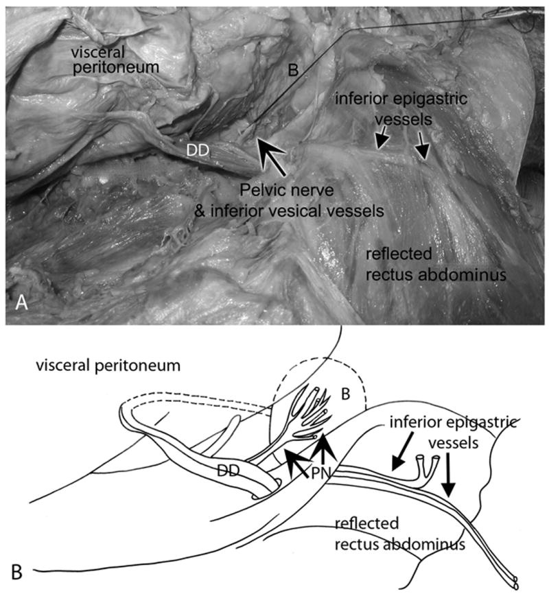
Figure 2.

Relationship of the pelvic nerve to the inferior vesical vessels, ductus deferens and inferior epigastric vessels. Lateral view; legs are located on the right side of these panels. The parietal peritoneum was separated manually from the visceral peritoneum and viscera. C) The vesical branch of the pelvic nerve (large arrow) was identified at the base of the bladder (B) in the retroperitoneal space. A suture was tied around the pelvic nerve for identification purposes. The suture is shown pulled inferiorly over the pubic bone (right side of panel). Pertinent landmarks such as the inferior epigastric artery and vein, ductus deferens (DD), and the reflected rectus abdominis muscle are indicated. (B) Diagram of the same figure showing the same landmarks. PN = pelvic nerve.
