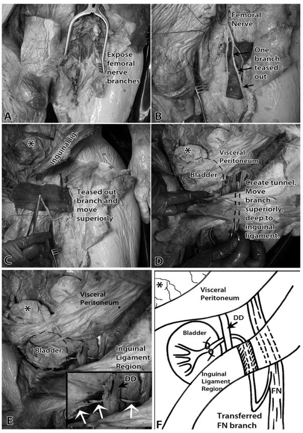
Figure 3.

Transfer of a muscular branch of the left femoral nerve to the left (ipsilateral) pelvic nerve. A) The branches of the left femoral nerve in the left anterior thigh were exposed. B) Branches of left femoral nerve were cleared of fat. A branch to the vastus medialis muscle was teased out from the femoral nerve trunk. A surgical cloth was placed under the nerve branches to aid visualization. C) This muscular branch was moved superiorly in the subcutaneous space. An asterisk indicates a loop of gut. D) The cut branch was tunneled inferior to the inguinal ligament into the abdominal cavity’s retroperitoneal space. The location of the bladder is indicated with a dashed line. E) The cut muscular branch of the left femoral nerve had enough length to be easily anastomosed with the left pelvic nerve on the bladder wall. Inset shows higher magnification of transferred femoral nerve and femoral-pelvic nerve “reanastomosis” site (white arrows). F) Diagram of image shown in panel E and of surgical procedure. Abbreviations: DD = ductus deferens; FN = Femoral nerve. The asterisk indicates the same loop of gut in each panel, as demarcated first in panel C.
