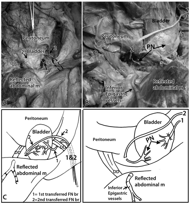
Figure 4.

Transfer of a muscular branch of the left femoral nerve to the right (contralateral) pelvic nerve. Anterior view, similar to Figure 3. A) Another muscular branch of the femoral nerve was identified and tunneled inferior to the inguinal ligament into the abdominal cavity’s retroperitoneal space. Tunnel is indicated by dashed lines. 1 = first transferred nerve (ipsilateral transfer); 2 = second transferred nerve (contralateral transfer). The left femoral nerve branch is moved across the abdomen, superior to the bladder, to the contralateral (right) pelvic nerve. B) Similar but enlarged image as shown in panel A. The cut muscular branch of the left femoral nerve (indicated with a 2) was placed adjacent to the transected right pelvic nerve (PN, left arrow). C) A diagram of panel A. D) A diagram of panel B. Abbreviations: DD = ductus deferens; m = muscle; PN = pelvic nerve; 1 = first transferred nerve (ipsilateral transfer); 2 = second transferred nerve (contralateral transfer).
