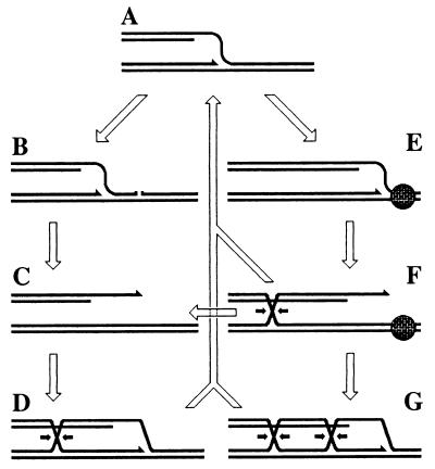
Figure 2.
The pathways of replication fork stalling/disintegration with subsequent resetting/repair. DNA duplexes are shown as double lines; a protein tightly bound to DNA is shown as a bricked circle. For all Holliday junctions, one of the two possible resolution directions is indicated by the small arrows. (A) A replication fork. (B) The replication fork approaching a single-strand interruption in template DNA. (C) The replication fork has collapsed at the interruption. (D) Double-strand end invasion to restore the replication fork structure. (E) A stalled replication fork. (F) Regression of the stalled replication fork forms a double-strand end and a Holliday junction. (G) Double-strand end invasion to restore the replication fork structure. Resolution of the Holliday junction in F leads to replication fork breakage (C). Resolution of the Holliday junctions in D or G, or exonucleolytic degradation of the linear tail in F leads to restoration of the replication fork structure (A).

