Fig 2.
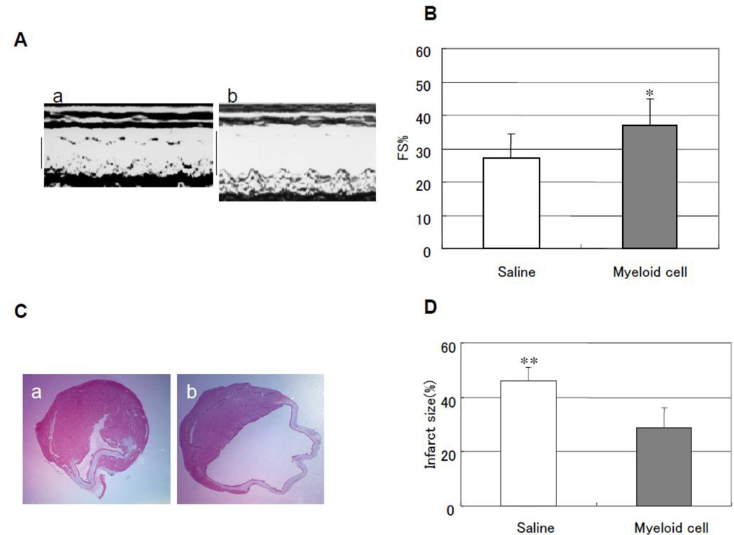
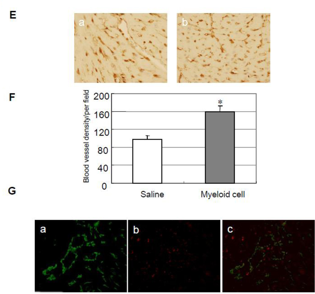
Intravenous injection of Gr-1+CD11b+ myeloid cells improves cardiac function and decreases cardiac infarct size after heart infarction via increasing neovascularization. (A) Representive echocardography of heart after 4 weeks of LAD ligation. a, Gr-1+CD11b+ myeloid cells injected from tail vain; b, saline injected from tail vain. (B) Gr-1+CD11b+ myeloid cells significantly improve FS after heart infarction compared with saline, *p < 0.05 versus saline, n=5 in each group. (C) Representive Masson's trichrome staining of the heart after 4 weeks of heart infarction, a, Gr-1+CD11b+ myeloid cells treatment; b, saline treatment. (D) Gr-1+CD11b+ myeloid cells significantly decrease cardiac infarct size after heart infarction compared with saline, **p < 0.01 versus saline, n=5 in each group. (E) Representive capillary density measurement in peri-infarct area of the heart. a, saline treatment; b, Gr-1+CD11b+ myeloid cells treatment. Magnification ×200. (F) Gr-1+CD11b+ myeloid cells significantly increase angiogenesis in the peri-infarct area after heart infarction, *p < 0.01 versus saline, n=5 in each group. (G) Florescent imaging of the heart after intravenous injection of Gr-1+CD11b+ myeloid cells. a, endothelial cells stained with anti-CD31 (green); b, Gr-1+CD11b+ myeloid cells marked with PKH-26 (red); c, some of the Gr-1+CD11b+ myeloid cells incorporate into vasculature (yellow). Magnification ×200.


