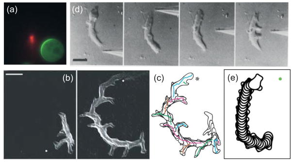
Figure 5. Response to chemoattractant gradients.

(a) Response of a latrunculin-treated Dictyostelium cell to a needle containing cy3-cAMP (red). The immobile cell forms a crescent of GFP-tagged PH-CRAC in the direction of highest concentration. (Reprinted with permission from Ref. 61. Copyright 2004 National Academy of Sciences USA.)(b,c) Overlays of a Dictyostelium cell chemotaxing to a cAMP-filled micropipette whose location (marked by the dot) is changed in the time between the two panels. The reorientation of the cell to the change in gradient, which is in the form of a gradual “u-turn” is seen in the overlay of the cell outlines in panel c. Scale bar is 20 μm. (Reprinted with permission from Ref. 28. Copyright 2007 Nature Publishing Group.)(d) Reorientation of a Dictyostelium cell by a sufficiently strong change in gradient. Here, the pipette is first moved to the back which stops cytoplasmic flow at which point applying the needle to the side leads to the emergence of multiple pseudopodia. Scale bar is 10 μm. (Reprinted with permission from Ref. 77. Copyright 1982 Elsevier Inc.)(e) Simulation showing the reorientation of a cell in response to a change in the direction of the chemoattractant gradient. (Reprinted with permission from Ref. 34.)
