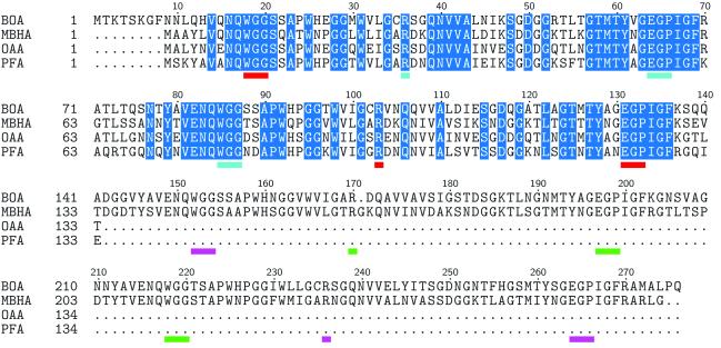
FIGURE 2.
Multiple sequence alignment of OAA family members. BOA and MBHA contain two sugar-binding domains, whereas OAA and PFA are single-domain proteins. Residues highlighted in blue are absolutely conserved among these lectins. Amino acids that interact with 3α6α-mannopentaose are underlined in red, cyan, magenta, and green, respectively, for binding sites I, II, III, and IV. Note that the binding site residues belong to the absolutely conserved subset in the alignment.

